Healing Heels: The Sole‑Searching Complexities of Heel Pressure Injuries
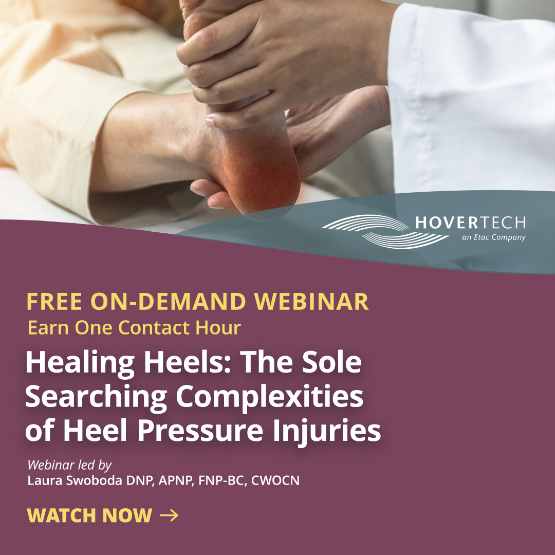
Pressure injuries of the heel are more common than many realize, accounting for 15 to 25% percent of total pressure injury cases. Like some pressure injuries, they’re painful and increase the risk of adverse outcomes and complications – and have added challenges of their own. Read one medical professional’s overview and recommendations and listen to the full on-demand webinar to earn one contact hour.
You’re familiar with pressure injuries: Tissue breakdown due to sustained, pressure-induced deformations, often exacerbated by environmental conditions. Pressure injuries are all too common across care environments, presenting most commonly in acute care.
Beyond the inherent risk they pose to patients (increased likelihood of sepsis, amputation, prolonged hospital stays, and death, to name a few), pressure injuries have implications for caregivers and healthcare, as well. They are quality care indicators used to evaluate the suitability of nursing care in healthcare settings. Even more, some 17,000 pressure injury related lawsuits are filed annually – the second most common claim after wrongful death.1
The good news: Once identified, pressure injury risk can be mitigated via use of suitable materials to disperse surface stresses and internal tissue stresses. This fact should galvanize prevention and treatment of one misunderstood pressure injury type in particular: pressure injury of the heel.
HEEL PRESSURE INJURY OVERVIEW
Pressure injuries typically occur in areas of the body where body weight is distributed over bony prominences: ankles, hips, shoulder blades, elbows, knees, ears, back of the head, and, yes, heels. Heel pressure injuries are more common than many realize. A recent 105,000 participant cross-sectional survey found that pressure injuries of the heel account for approximately 18.2% of total pressure injury cases. A separate cross-sectional survey of 85,000 individuals found prevalence to be even higher – between 23% and 26%.2 The takeaway: At minimum, nearly one in five individuals with pressure injuries will experience them on one or both heels. And when it comes to deep tissue pressure injuries, the heel is the most common site. An astounding 41% of all deep tissue injuries occur on heels.
Meanwhile, heel pressure injuries are commonly misdiagnosed and overlooked. The prevalence of deep tissue injuries on the heel is partly a reflection of such oversight. Misdiagnosis and oversight occur across skin tones but are especially prevalent among darker skin tones. Consequently, heel pressure injuries among patients with darker skin tones are commonly more progressed upon diagnosis, sometimes failing to be recognized until reaching Stage 3 or Stage 4.
Pressure injuries of the heel introduce risks to the patient inherent to any pressure injuries, and a few more: Their placement on the heel can reduce mobility and make patients non-ambulatory, contributing to cardiovascular deconditioning that can lead to an increased incidence of cardiovascular mortality. And for a variety of reasons including delayed treatment, research shows a strong positive correlation between the development of heel pressure injuries and increased mortality rates as well as increased litigation.
Put another way, heel pressure injuries are a significant issue, especially given the high incidence of peripheral artery disease among patients who suffer from them (one study found peripheral arterial disease in 73.5% of heel pressure injury cases).3 And treatment of heel pressure injuries is complicated. The heel of the foot is comprised of uniquely thick tissue that often defies reconstruction. The added layer of skin on the heel is called the stratum lucidum. It’s largely protective, but difficult to reconstruct – and one of many unique aspects of heel anatomy and physiology.
ANATOMY AND PHYSIOLOGY OF THE HEEL: IT’S COMPLICATED
Because they’re difficult to heal, many assume heels are not well vascularized. In fact, the opposite is true. Heels are richly vascularized by three arteries –the posterior tibial, sinus tarsi, and peroneal (sometimes called the fibular artery). The challenges of healing heels are byproducts of other anatomical and physiologic factors:
Fascia. The thick band of tissue that connects the heel bone to the toes, fascia is poorly vascularized. When injured or otherwise interrupted, permanent deficits can develop that lead to protracted healing and/or pain moving forward.
Bony prominence. Limited amounts of tissue beneath the calcaneus bone (or heel bone) translate to limited support and padding, making the area uniquely susceptible to the effects of pressure.
Loss of heel fat pad. The heel fat pad covers and therefore protects the vulnerable fascia and calcaneum bone by absorbing and transferring the impact of walking (of note: walking puts force two and a half times a person’s body weight on the heel, one of many reasons doctors might recommend weight loss). The heel pad fat is the only hope against heel pressure injuries for the poorly vascularized fascia in particular – so when it’s diseased, or lacking in cushion and elasticity, pressure injury risk grows exponentially. Risk factors for heel fat pad syndrome include age, prolonged standing, high impact activities, insufficient footwear, and obesity.
Now consider the plethora of medically related risk factors for heel pressure injury:
- Immobility
- Diabetes
- Vascular disease including peripheral artery disease (PAD)
- Impaired nutrition
- Perfusion issues
- Mechanical ventilation
- Surgery, perioperative, or angiosome type of injury to the heel
- Braden subscales
Even more, patients who are hospitalized, have chronic diseases, and/or reside in long-term care or assisted facilities tend to be positioned in one of three ways: supine position, Semi-Fowler’s, or Fowler’s. In each of these, the heel typically makes contact with the bed, applying pressure over a bony prominence and, often, cumulative pressure over time, collectively increasing risk of heel pressure injury.
Patients in critical care settings are especially vulnerable, for the reasons above as well as the prevalence of circumstances that diminish tissue tolerance, which is the ability of skin and its supporting structure to endure pressure without adverse sequelae:
- Increased moisture (fever, incontinence, sweating including diaphoresis)
- Medical devices, which often make physical contact with skin
- Low oxygenation and other heightened vascular impairments
- Risk of the COVID virus, which impairs the inner lining of blood vessels and directly infects the vascular endothelium
INDIRECT PRESSURE INJURY VIA ANGIOSOMES
A realization that’s newer to some segments of the wound community is that pressure injuries can develop indirectly, away from the site where pressure was applied. Dr. Caroline Fife brought this to light after noticing that her son developed heel pressure injuries post-operatively even though pressure from a surface pillow was on his calves, not his heels.4
We now understand why: The pressure from the pillow occluded one of the arteries more proximally. Tissue death subsequently occurred over the heel because its artery supply was cut off.
Angiosome is the term for that whole wedge of tissue that’s supplied, or perfused, by an artery.
Occlusion of a named vessel supplying that angiosome, which can occur via external pressure, poses the risk that that whole wedge of tissue then dies. Maps of the body exist showing the placement of angiosomes, some of which are in the foot.
This type of pressure injury tends to be perioperative, resulting from positioning during an operative procedure or during imaging. It can also present among patients who undergo CT or MRI scans repeatedly, or who undergo dialysis and are therefore in a positioned state for extended periods of time.
The takeaway: using pillows and alternative positioning devices that aren’t designed to offload the heel can contribute to some of these angiosome-based heel pressure injuries.
HEEL PRESSURE INJURY INTERVENTIONS
The billion-dollar question *holds pinky to mouth*: what can be done to prevent pressure injuries of the heel?
Recommendations below are an amalgamation of NPIAP (The National Pressure Injury Advisory Panel) new “living” guidelines as well as expert consensus among pressure injury prevention professionals and researchers:
- Moist wound healing. Betadine alone is rarely sufficient. Covering a wound with protective dressing maintains a moist wound environment, minimizes bacterial contamination, maintains normothermia, and protects the wound from trauma. However, moist wound healing is not a panacea, i.e. is not suitable for every patient at every point. Do not apply moisture to stable, non-infected ischemic heel ulcers – pressure injuries stemming from severe peripheral artery disease, whereby patients lack sufficient blood flow to heal and are being protected by eschar. Wounds that are stable, dry, non-infected and profoundly ischemic should be left in place.
- Prophylactic dressing. In one study of high-risk intensive care unit patients, after the sacral dressing began being used, the number of unit-acquired sacral pressure ulcers decreased by 3.4 to 7.6 per 1000 patient days depending on the unit.5 These results indicate that the application of dressings in high-risk intensive care unit patients is effective in preventing pressure ulcers in these areas of the body. Prophylactic dressing comes in many forms: multi-layer foam, multi-layer super-absorbent polymer, hydrocolloids, and hydrogels for pressure injury prophylaxis.
- Manual repositioning and heel elevation. These are perennial and proven ways to offload heel pressure and reduce risk of pressure injury. Ideally, elevated heels are fully free from the support surface.
- Offloading boots and devices. Available in both ambulatory and non-ambulatory, offloading boots are specifically designed to envelop and support tissue and dissipate force over a wider surface area beyond the bony calcaneal prominence. Like moist wound healing, offloading devices aren’t universally recommended. A heel offloading device needs to be appropriate for the patient’s mobility and activity level. When choosing from offloading options, consider patient preference, anatomical differences (including any altered bony prominences), skin integrity, edema, levels of activity and mobility (remember that boots are a fall risk), and clinical condition and tolerance (agitations, muscle spasms, kicking, scooting, etc.).
- Repositioning wedges. Pillows, gloves, and filled saline bags can also be used for repositioning if needed – again, being sensitive to the risk of pressure injuries via angiosomes. Clinically approved options are always best.
In terms of what to avoid: Sheepskin (even medical-grade sheepskin) is not shown to effectively offload pressure from the heels or to reduce the occurrence of heel pressure injuries. This reality is reflected in recently updated NPIAP guidelines. However, since sheepskin has been used for years, if a patient doesn’t want to cease use despite recent evidence, meet this patient where they are.
Lastly, a quick note on whether prophylactic dressing and offloading boots should be used simultaneously – a question that comes up often. There’s no clinical evidence pointing to the value of using both interventions at the same time.
With that said, clinicians, specialists, and certified wound specialists who know patients best should feel empowered to make the call, as it sometimes feels like the best choice to use both. Remember that patient tolerance and sensitivity should factor into decision-making.
OTHER CONSIDERATIONS
It’s easy to list out interventions. It’s another to effectively fold them into dynamic healthcare environments full of competing priorities. Below are additional actions correlated with more successful pressure injury treatment outcomes overall:
- Offloading and repositioning devices that are accessible and stocked locally, close to the point of care, are more likely to be initiated in a timely manner.
- Organizational methods that support the use of offloading devices for heel pressure injuries also lead to more and better use. These include established protocols for stocking and supply, training and education, even auditing.
- Proactive encouragement and involvement from leadership and management can also increase use and positive clinical outcomes.
Of course, this is just the tip of the iceberg. Learn more about heel pressure injury causes, treatment, and prevention and earn one free contact hour by listening to the full on-demand webinar.
Laura Swoboda, DNP, APNP, FNP-C, FNP-BC, CWOCN-AP, WOCNF, CWCP

A translational scientist, family nurse practitioner, and practicing skin and wound specialist in Milwaukee, Wisconsin, Dr. Swoboda serves as a principal investigator for quality improvement, evidence-based practice, and research projects in complex skin and wound care.
Dr. Swoboda is president elect of the Milwaukee Metro Nurse Practitioners Association. They serve as treasurer for the AAWC, and on the board of directors for the Save a Leg, Save a Life Foundation. Dr. Swoboda also serves on various committees for the NPIAP, Wound Healing Society, WCCC, Alliance of Wound Care Stakeholders, and the American Heart Association’s Peripheral Arterial Disease Collaborative. Dr. Swoboda is committed to health equity. They volunteer at the local, regional, national, and international levels to increase access to quality healthcare.
- https://www.ahrq.gov/patient-safety/settings/hospital/resource/pressureulcer/tool/pu1.html
- https://pmc.ncbi.nlm.nih.gov/articles/PMC7155923/
- https://www.researchgate.net/publication/359743335_Influence_of_Peripheral_Arterial_Disease_on_Wound_Healing_in_Heel_Pressure_Ulcers#:~:text=Abstract,PAD%20are%20difficult%20to%20treat.
- https://carolinefifemd.com/2024/10/14/severe-pressure-ulcer-malpractice/
- https://aacnjournals.org/ajcconline/article-abstract/25/3/228/3143/Prophylactic-Sacral-Dressing-for-Pressure-Ulcer?redirectedFrom=fulltext
