Skin Injury’s Two Risk Phases: Understanding and Preventing Pressure Injury Across the Continuum of Care
An estimated 2.5 million U.S. patients develop hospital-acquired pressure injuries (HAPI) annually, in part because of lack of awareness around causes and related symptoms. Below, we’ll shed light on skin’s two distinct risk phases and offer practical, evidence-based HAPI prevention tactics for each, applicable to every shift in every unit. Listen to the full on-demand webinar to earn one contact hour.
Despite growing awareness, pressure injuries continue to take a catastrophic toll on human lives. An estimated 60,000 patients in the U.S. die annually from pressure injuries and related complications
And more than lives are on the line. Pressure injuries cost the U.S. healthcare system $26.8 billion annually. Stage 3, Stage 4, and unstageable HAPIs are designated never-events by the Centers for Medicare & Medicaid Services, meaning healthcare organizations are not eligible for reimbursement if these wounds develop under their care.
The onus is on healthcare providers to prevent pressure injuries, which can develop in any inpatient or specialized setting. As any healthcare professional knows, this is easier said than done. However, preventing HAPIs is not impossible. In fact, you might be surprised to learn that ninety-five percent of HAPI are considered preventable.
Why, then, do HAPI persist? is the next logical question.
The answer touches on many systemic challenges ranging from competing priorities facing practitioners to the interdepartmental collaboration required to head off HAPI. However, there is one relatively low-barrier way to help keep HAPI’s at bay: Educate practitioners on the mechanisms that cause them. While most pressure injuries indicate a breakdown of skin and tissues, the exact drivers of these skin injuries can vary dramatically. When understood and woven into caregiving, small, right-fit choices can go a long way toward prevention. Below we’ll review the two primary skin risk phases for pressure injury development: Movement and Dwell Time, after a discussion on skin vulnerability.
Understanding their subcategories – shear, friction, and pressure – will lead to an introduction of evidence-based prevention strategies for each. When you reach the end of this piece, you’ll hopefully have a mental HAPI prevention framework you can use in every patient interaction.
WHY SKIN IS VULNERABLE: A QUICK REVIEW
Remembering when and why skin is vulnerable requires a quick, foundational anatomy and physiology review:
Skin is the body’s largest organ and accounts for 16% of the entire human body. It’s comprised of three layers:
- Epidermis – the top/outer protective layer of skin that regenerates every two to four weeks.
- Dermis – the layer beneath the epidermis, which contains collagen, elastin, blood vessels, and nerve endings.
- Hypodermis – the subcutaneous layer, containing fat, connective tissue, and other protective elements that cushion the bone
Skin is at risk when tissue breaks down. The reason this can occur:
- Pressure above 32 millimeters of mercury (mmHg) can block skin’s top layer blood flow, occlusion. Illustrating how easily occlusion can occur: Simply laying on a standard mattress can generate anywhere from 60 to 150 mmHG of pressure over bony prominences. Bending and kinking of blood vessels can also contribute. Occlusion leads to ischemia (restricted or reduced supply of blood flow to the skin), which can lead to anaerobic metabolism and cell death.
- Muscle at skin’s deeper layers is uniquely vulnerable to ischemia, sometimes occurring independently of outer later damage.
TWO SKIN RISK PHASES: MOVEMENT AND DWELL TIME
This leads to discussion of the two distinct risk phases for skin – movement and dwell time:
- Movement is anytime a patient is moved, including transferred, boosted, turned, repositioned, or otherwise handled by clinicians.
- Dwell time is any time a patient is not moving, regardless of their position.
Understanding the difference is key to effective preventive care. Because while movement and dwell time can both cause pressure injuries, their clinical drivers are very different:
- The clinical drivers during movement are friction and shear.
- The clinical driver during dwell time is sustained pressure.
- Sustained pressure from medical devices – a different form of dwell time – can also lead to pressure injury
It’s important to remember that all patients experience both movement and dwell time on any given day. Consider a critical care patient. Their skin is susceptible during standard repositioning (movement) and while lying still the remainder of the time between turns (dwell time). This is important to note because the combined impact of movement and dwell time is often more damaging than the sum of its parts. The two work in tandem to compound skin damage, proving more detrimental than one or the other alone. Clinically, this means practitioners must be aware of and ongoingly working to minimize the impact of both risk phases.
It starts by understanding their mechanical differences.
MORE ABOUT SKIN RISK PHASE 1: MOVEMENT
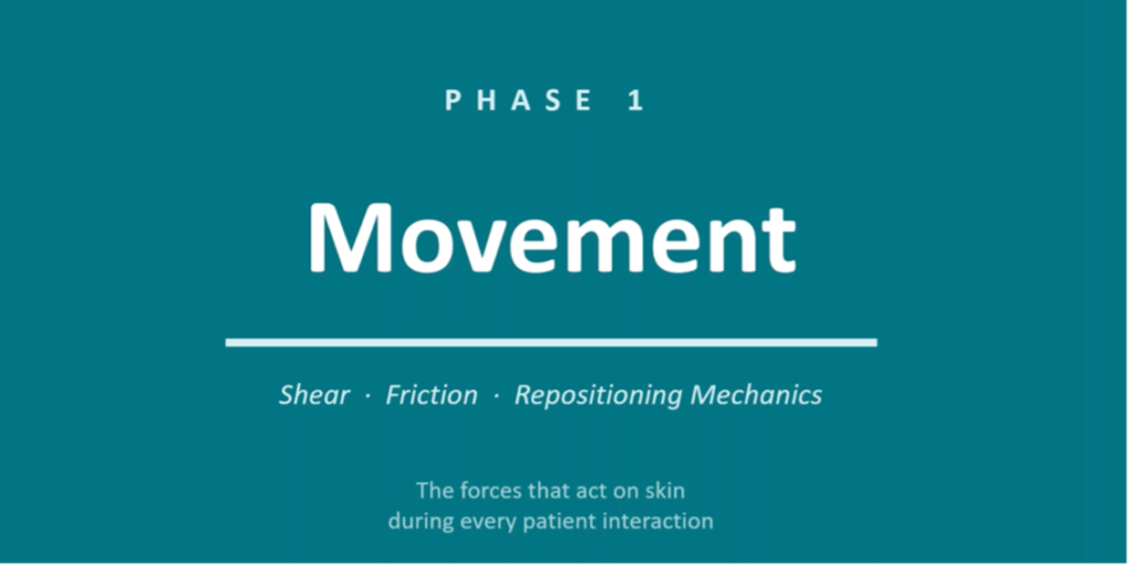
Shear and friction are the two mechanical forces introduced during patient movement. Every time you handle a patient, shear and friction to some extent take place.
These, too, are distinctive from one another:
Shear
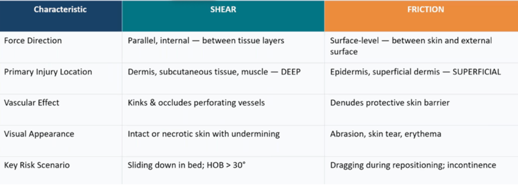
Sometimes called the hidden killer of tissue, shear occurs when layers of skin tissue slide in opposite directions relative to one another, bending and kinking the blood vessels that supply the dermis and subcutaneous tissue – a process known as angulation. Angulation results in occlusion, which damages the deeper layers of the skin: the dermis, hypodermis, fat, and even muscle.
Because this injury occurs in the skin’s deeper layers, resulting pressure wounds are not always easily seen. Extensive blood vessel damage to the sacrum caused by shear, for example, can look like a mere bruise on the surface.
Many common clinical scenarios create shear. The most pervasive is when a patient slides down in their bed, with their skin against the bedsheet. The patient’s skin stays in place against the sheet while deeper tissues shift downward with the body’s skeleton. Blood vessels consequently start to stretch and kink.
Other common clinical scenarios that present a high risk of shear:
- Head of bed (HOB) elevated/tilted at an angle greater than 30°
- Pulling a patient up in bed repeatedly
- Semi-Fowler’s position
- Sliding transfers (bed to stretcher and back, for example)
- Positioning an OR table
- Scooting across surfaces instead of lifting
Friction
Friction is resistance force between two surfaces moving across or rubbing one another (when skin is dragged across a coarse surface), impacting the outermost epidermis. Repeated abrasion denudes the skin’s primary barrier, creating superficial wounds that become entry points for bacteria. Unlike shear which causes deep internal vascular damage, friction causes surface-level damage.
Friction, too, is pervasive in clinical settings. Picture a nurse pulling a patient up in bed. Even if using a glide sheet, the patients’ back and heels are going to drag. That dragging can fully abrade the epidermis.
Friction is exacerbated by other common risk factors, such as incontinence. Macerated skin has a 5 times higher friction coefficient. If skin and/or sheets are damp, skin is already macerated, making it much more susceptible to friction during movement. Skin fragility is another risk factor. Elderly, corticosteroid-thinned, or edematous skin tears much more easily under pressure. Speed of movement also comes into play. Rapid movement, like fast repositioning, increases friction force. Also increasing risk is the fact that friction injury can develop fast – in less than 30 minutes.
Some preventative tactics specific to reducing friction:
- Apply barrier creams or films to at-risk skin (most commonly, the skin on the elbows, heels, and sacrum)
- Dress wounds and fragile skin before repositioning rather than after
- Reposition and otherwise handle patients via slow, supported movement
To help you remember the differences between friction and shear, here is a clinical comparison of the two across five characteristics:
SAFE REPOSITIONING: REDUCING PHASE 1 FORCES
As mentioned, patient movement is inevitable. How, then, do healthcare providers decrease the chances of friction and shear? How do we reduce these forces? It’s the little repetitive clinical choices, after all, that can initiate or prevent skin damage.
This practical checklist is where to start. Keep in mind that these rule-of-thumb tips are not suitable for every scenario. They are patient dependent and should only be utilized when clinically appropriate:
- Lower head of bed (HOB) to flat. Nurses and other practitioners sometimes don’t realize that elevating the HOB higher than a 30° angle dramatically increases the chance of shear. Briefly lower the HOB to flat (or to less than 10° angle) during repositioning, when risk is highest-if the patient’s status allows. Restore the bed to the prescribed elevation after.
- Use a friction-reducing device. Air assistive devices can reduce the risk of friction by up to an astounding 80%.
- Minimum 2-person technique. Never move a patient without at least two practitioners involved. One person is stabilizing the patient while the other positions. This especially helps prevent dragging and shear during lateral turns.
- Lift, don’t drag. When moving a patient, be sure to clear the surface with their body instead of dragging it. This reduces cumulative skin force exposure. Sometimes this means enlisting more than two people and/or the help of mechanical or ceiling lifts.
- Protect heels during transfers. Heel-based pressure injuries are the second most common, after sacral injuries, in large part because heels tend to be dragged during bed transfers. Heel pressure injuries can be hard to notice or spot because of where they’re located. They’re also difficult to treat, making prevention incredibly important – especially during patient repositioning.. Use heel protectors or elevate heels during every turn.
- Assess skin immediately post-move. Once you’ve repositioned a patient and confirmed they’re comfortable, closely assess the skin. Check the sacrum, heels, elbows, and occiput after every reposition, and document findings (more on documentation below).
MORE ABOUT SKIN RISK PHASE 2: DWELL TIME
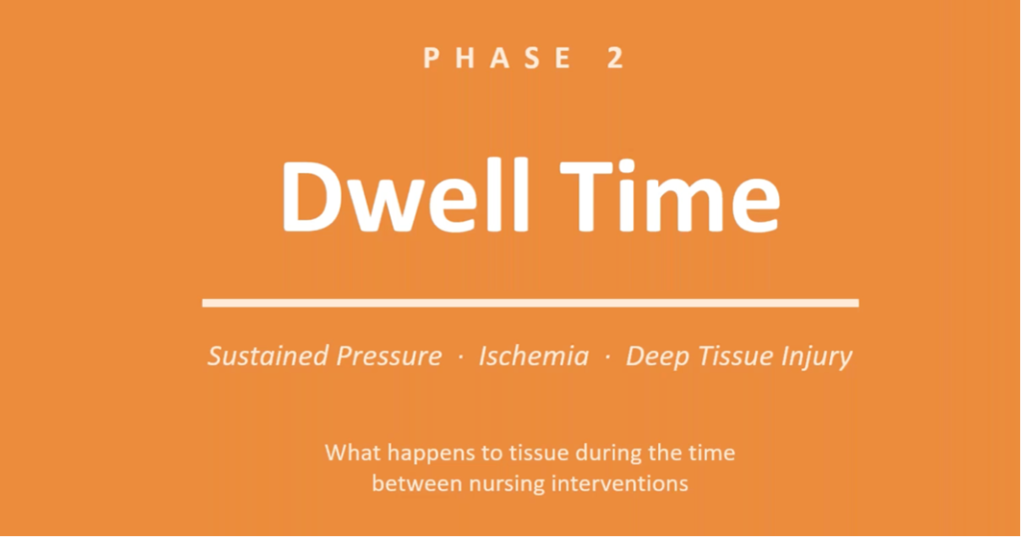
The second of skin’s two risk phases occurs anytime a patient is not moving. ICU/critical care patients experience dwell time most frequently, as Q2 turning is sometimes the only opportunity for movement.
The clinical driver of skin injury during dwell time is sustained pressure, leading to ischemia that can lead to deep tissue injury. Because pressure from dwell time affects skin’s deeper layers, it sometimes progresses to become a deep tissue pressure injury (DPTI), which can progress to Stage 3, Stage 4, or unstageable pressure injuries in as little as three days.
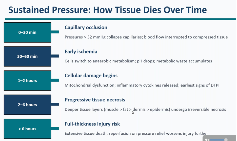
This timeline of tissue death shows how occlusion can cause irreversible tissue death within six hours:
From a skin risk perspective, dwell time is just as dangerous as phase one movement. It’s similarly pervasive across patient populations. Here is a descending list of high-risk populations and the reasons they might be vulnerable:
- Critical Care Patients – vasopressors, sedation, multiple medical devices, hemodynamic instability, chronic immobility. In the absence of aggressive prevention efforts, HAPI rates among critical care patients can reach as high as 30%.
- Perioperative Patients – hypothermia, vasoconstriction, prolonged time on an operating room’s hard surface, hypothermia. Perioperative conditions can jeopardize intact skin in an alarmingly short amount of time.
- Spinal cord injury patients – loss of sensation (including pain response), loss of voluntary movement.
- Elderly and long-term care (LTC) facility patients – thin and fragile skin, malnutrition, incontinence, cognitive impairment, reduced mobility, infection risk.
- Bariatric patients – skin folds that can trap moisture, skin-to-skin contact in skin folds, limited repositioning options, underperformance of or incompatibility of standard prevention equipment (wedges, offloading devices) in light of patient size and weight. Bariatric patients are a special population. It is essential to check their skin folds, perform assessments, and ensure that your facility is prepared to take care of these patients’ skin in terms of awareness, care, methods, and equipment (bariatric beds and mattresses, for example).
- Palliative/end-of-life patients – rapidly declining skin perfusion, repositioning that conflicts with comfort goals. End-of-life patients are another special population sometimes necessitating breaks from protocol (repositioning frequency is sometimes decreased, for example, based on the patient and family decisions). Note that some unique pressure injuries that develop at end-of-life are the result of the dying process, not preventative care failures (chief among these, the Kennedy Terminal Ulcer).
Because deeper skin injuries can be difficult to spot, thorough skin assessments are key. This is especially true of deep tissue pressure injuries. Because DTPI develops deep in the tissue and works upward, by the time it’s visible at the surface, damage is already being done. Here are ways to assess for DTPI and deeper pressure injury development:
Keep in mind: DPTI development is often indicated by deep red, maroon, or purple skin. However, this discoloration can be difficult to see on darkly pigmented skin, underscoring the importance of tactile assessments.
Perform a tactile assessment. Be on the lookout for skin that is uniquely firm, boggy/indurated, warmer or cooler than surrounding tissue, or painful for the patient.
OFFLOADING PRESSURE: REDUCING PHASE 2 FORCES
Q2 turning – the medical protocol in which a bed-bound patient is repositioned every two hours at minimum – is a proactive measure designed to help prevent pressure-induced skin injury. But there are several more proactive and tools at practitioners’ disposal:
- The 30° lateral tilt. Why a 30° angle? Why not 90°? Wouldn’t 90° take more pressure off the patient? This is a common misconception. Tilting patients to 30° in bed is often the optimal choice for several reasons:
- Unlike a 90° lateral tilt, which concentrates pressure directly over the greater trochanter (a bony prominence with limited soft tissue padding), a 30° tilt puts more pressure over the fleshy lateral hip/gluteal area, ensuring no bony prominences are directly loaded.
- The 30° tilt can generally be tolerated better than 90° by frail, elderly, and/or post-operative patients.
- Wedges. Typically made of foam, wedges help patients maintain desired positions including the 30° tilt. Here are some practical applications of wedge-supported options:
- Sacrum/coccyx – Aim for that 30° lateral tilt with the wedge(s) under the hip and back. This next part is imperative: Make sure the sacrum is clearing the mattress surface, (i.e., is free floating). Avoid placing the wedge directly under the sacrum. After the wedge is placed and the patient is tilted back, use your hand to verify that the sacrum is still free-floating. This is a common oversight.
- Heels – As the second most common site for pressure injuries, heels deserve special emphasis and care. One of the reasons heels so commonly develop pressure injuries is the standard action of putting a pillow under the heels or legs to offload calves places heels into direct contact with the pillow surface. Sometimes the only appropriate heel offloading is total suspension, freeing the heels of any contact with a pillow or other surfaces. Place a pillow lengthwise under the calf to suspend the heel completely off the mattress. There’s a little mnemonic that can help you remember this: FLOAT the heel – fully lift it off and touch the heel. Flex the knee slightly or use a heel offloading boot to avoid popliteal pressure. If using a boot, check the Achilles tendon for pressure from the boot at every assessment.
- Occiput/ears – The occiput (area behind the ears and base of the skull) is a common site of medical device pressure injuries. Each device presents its own offloading challenge, but as a rule of thumb: reposition the head every two hours and use a gel pad or a donut-free head positioning device (donut cushions increase perimeter pressure and should therefore be avoided).
- Elbows/lateral malleolus – Place soft foam protectors or pillows under elbows and between knees for lateral-lying patients, as lateral malleolus (the bony knob on the outside of the ankle) is a risk when patients are positioned in the 30° lateral tilt. Check skin under all devices at each assessment.
Of course, no single intervention is enough to prevent pressure injuries. Pressure injury prevention in dwell time circumstances is proactive action from every single angle. Luckily, bundles exist for this purpose. More on those below. But first, a look at variations in patients’ susceptibility to skin injury.
TISSUE TOLERANCE: WHY SOME PATIENTS INJURE FASTER
Not all patients are equally vulnerable to pressure injuries. Two patients under identical pressure conditions may have completely different skin profiles and outcomes based on their underlying physiology. Boiled down, these disparities stem from differences in tissue tolerance – the ability to withstand pressure and deformation – which is determined by multiple intrinsic and extrinsic factors:
Perfusion & oxygenation
- Peripheral vascular disease – this is the most critical modifiable fact dictating skin vulnerability to ischemic injury resulting from reduction in continuous blood flow, even at lower pressures. Common among patients on vasopressors, norepinephrine, and norepinephrine, which reduce continuous blood flow.
- Hypotension/vasopressor use
- Anemia (Hgb<8 g/dL)
- Diabetes mellitus
Nutrition status – matters more to wound care and wound healing than is commonly recognized.
- Malnutrition/hypoalbuminemia
- Serum albumin < 3.5 g/dL
- Dehydration
- Low BMI
Skin & tissue condition
- Patients with edema have two complications: mechanical vulnerability (stretching of the skin, making it more fragile) and microvascular vulnerability (fluid in their interstitial space that impairs oxygen diffusion)
- Dry skin
- Prior pressure injury scars
- Corticosteroid thinning
Neurological factors – these can impact the ability move, the ability to know when to move, and ability to communicate
- Spinal cord injury
- Sensory neuropathy
- Sedation/paralysis
- Altered consciousness
A patient that meets three or all four tissue tolerance deficit categories above is at very high risk of pressure injury development (ICU patients often check all four boxes). The critical implication here is that these patients are going to need more frequent repositioning and more intensive prevention strategies than your standard patient. Specifics will be determined by the Braden Scale.
THE VALUE AND ROLE OF THE BRADEN SCALE
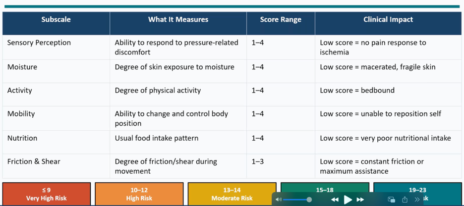
Specifics of patient skin care should be guided by the Braden Scale, the most widely used and validated pressure injury risk assessment tool in medicine.
The Braden Scale gives practitioners an opportunity to enter a patient score, ranging from 1 to 4, across subscales including moisture, sensory perception, and, yes, friction and shear. The score drives the specifics of intervention. A patient with a low sensory perception score who can’t self-report requires more frequent skin assessments per the Braden Scale, for example. A low moisture score will drive more frequent and specific incontinence care protocols, such as barrier treatments.
The Braden Scale is designed to be completed within eight hours of patient admission (or per your facility’s policy) and repeated with any significant change in the patient condition. Like all standardized assessments, it’s not without its limits. Because it was developed primarily alongside a Caucasian population, research suggests that the Braden Scale might under-predict skin injury risk in patients with darker skin tones. This sobering fact leads to a critical reminder: the Braden Scale and all formulas should always be supplemented by clinical judgment. Note, too, that a modified Braden Scale exists for pediatric patient populations.
PRESSURE INJURY PREVENTION BUNDLES
Discussion of varying tissue tolerance and different patient populations naturally leads to discussion of the pressure injury prevention bundle.
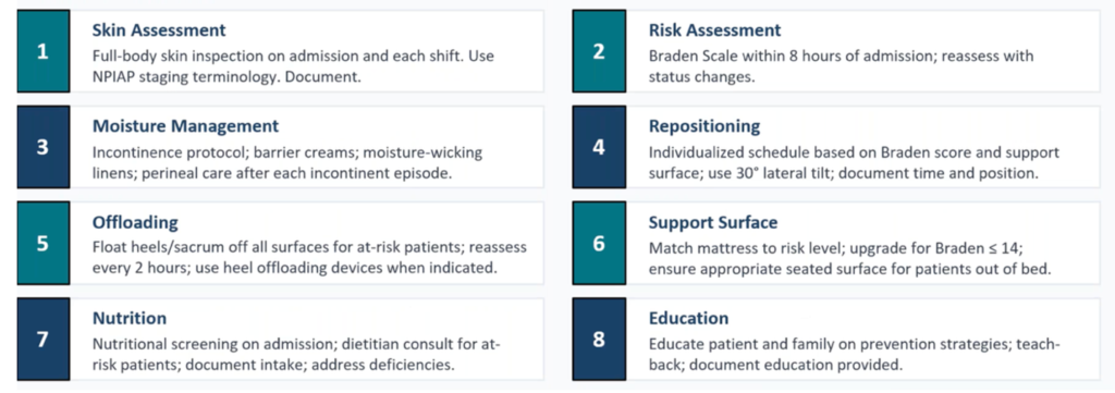
Bundles are built on the earlier-stated fact that no single intervention alone is as effective as multiple interventions. A carefully devised intervention suite, when thoughtfully implemented within a time frame, is your best bet at protecting skin integrity. Utilizing a bundle greatly improves healthcare outcomes.
Here is a glimpse at the Agency for Healthcare Research and Quality (AHRQ) Prevention Toolkit bundle:
SKIN INJURY PREVENTION ACROSS THE CARE CONTINUUM
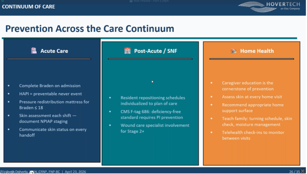
So what does skin injury prevention look like across the continuum of care, from admission through discharge and beyond?
Below is a go-to overview of skin considerations and related actions across a patient journey, from acute care to home health. Again, clinical specifics might dictate patient-to-patient modifications.
Keep in mind, too, that home health care often is not overseen by professional caregivers but by a patient’s family and personal support team. Before a patient is discharged or continues on to their next step, then, nurses should consider having a teach back session with family on things they might not know or understand: Q2 turning, repositioning, moisture management, and skin checking, to name a few. What should they be looking for while assessing skin? What should worry them, what’s normal? What should cause them to seek added support? Use care in efforts to close that education gap.
DOCUMENTATION, COMMUNICATION, AND OTHER RECOMMENDATIONS
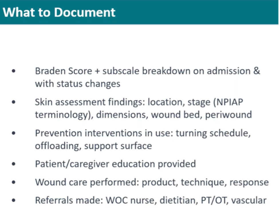
- Document, document, document. The critical importance of documentation in skin care cannot be overstated, for all reasons including legal ones. A Stage 2 pressure injury that progresses to Stage 4 is going to raise serious legal and quality of care questions. Observations around skin and any existing wounds should therefore be documented at every care setting. When documenting, use precise language and approved National Pressure Injury Advisory Panel (NPIAP) terms that align with professional standards: Stage 1, Stage 2, and unstageable, to name a few. These terms are far more useful and airtight than skin discoloration notes. If your facility allows it and if it’s in your workflow, timestamped, well-lit photographs can be incredibly beneficial. Documentation in end-of-life care should be even more thorough than standard skin documentation, establishing end-of-life goals as determined alongside a patient’s family and using it as rationale for care offered, which needs to be outlined in detail:
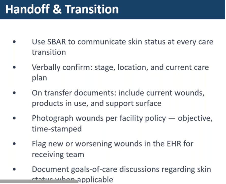
- Prevent handoff gaps. Research shows that skin-related inflammation is commonly omitted from data shared during patient handoffs and transitions, such as shift changes and transfers. Make sure the care teams you’re working with create a habit of including skin status at every handoff, with every patient – even those the patient currently has no wounds. Remember, a patient with a perfect skin assessment today could already have a festering injury waiting to present itself in days ahead.
- Assign a skin champion. Keep skin top of mind by designating one or two nurses per unit who initiate and lead daily pressure injury prevention conversations. Skin champions let teams know when there’s new research or literature, and what’s working, what’s not. Ideally, these skin-related conversations are woven into daily huddles. Skin champions can round on high-risk patients, mentor new nurses, and all in all keep pressure injury prevention at the forefront of consideration and patient care.
- Celebrate the wins. Last but not least, be sure to acknowledge and celebrate when a unit has zero pressure injuries. Acknowledging these moments is encourages the teams working incredibly hard, day in and day out, to keep skin intact against extremely challenging circumstances.
Eager to learn more about preventing hospital-acquired pressure injuries? Earn one free contact hour by listening to the full on-demand webinar..
Elizabeth Doherty, MSN, CRNP, FNP-BC

Elizabeth brings a broad range of clinical expertise to her role as Clinical Education & Training Manager at HoverTech International. Her background spans long-term care, medical-surgical ICU experience at a large academic hospital, emergency and pediatric emergency nursing in a Level I trauma center, and advanced practice as a Certified Registered Nurse Practitioner.
This diverse foundation allows her to connect with healthcare teams across multiple specialties and understand their unique challenges in patient care and safety.
She is deeply passionate about shaping the future of nursing, supporting frontline nurses, and ensuring they have the knowledge, tools, and confidence to deliver safe, high-quality care.
